Guided Implant Surgery
At Dream Dental, we invest in the latest technology available in dentistry today, to provide the highest level of care to our patients.
Latest Technology in Dentistry
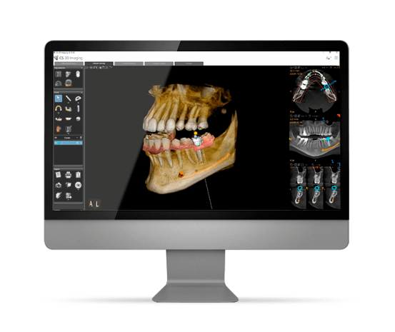
Guided implant surgery starts with a meticulous and highly detailed planning process and requires the use of the latest imaging technology in medicine. First we take multiple images of your mouth with a special intra-oral camera and a CBCT scanner (3D scanner) to be able to study your teeth and surrounding oral structures.
Surgical Simulation Before Treatment
We use a sophisticated surgical simulation and treatment planning software to predict the outcome of your treatment even before actually performing the surgery. With the help of this amazing technology, we are able plan out the entire surgery step-by-step before your procedure begins. Then, based on the digital information from the surgical simulation, our dental laboratory creates a precise surgical guide.
Precision with Surgical Guide
By the time of your procedure, your surgeon will already be familiar with the details of important anatomical landmarks in your mouth. The surgical guide will enable your doctor to place the implant in a precise location, depth, and angle.
No Incisions, No Sutures
Because your surgeon already knows the details of the surgical site, there is typically no need for an incision of the gum tissue, and no sutures would be required. The most important benefits of this implant surgical technique are the extremely low risk of complications and significantly shortened healing time.

CS 8100 3D
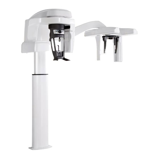
The CS 8100 3D delivers outstanding image clarity in all modalities with its latest enhancements. This simple 4-in-1 solution offers excellent value and no compromise on quality.
- Versatile 2D/3D System. The CS 8100 3D blends 2D panoramic technology and CBCT imaging with 3D model scanning to create one powerful unit.
- Selectable Fields of View. It supports all diagnostic needs while limiting patient exposure and area of responsibility. Smart, laser-free face-to-face positioning facilitates proper patient placement and increases accuracy.
- High-Resolution 3D Images & Noise-Free The CS 8100 3D delivers ultra-high-resolution images. Meanwhile, the ANR algorithm reduces image noise while preserving clinical details.
- Plan Implants with Confidence. Combining accurate 3D images with intuitive implant planning software, the CS 8100 3D is an ideal solution for confidently placing implants.
- Clear & Sharp Panoramic Images. The new Tomosharp algorithm is combined with the latest image processing technology to deliver ultra-sharp panoramic images instantly.
- Metal Artifact Reduction Technology. CS MAR1 drastically reduces metal artifacts caused by dental restorations, implants, and fillings. This helps Dr. Khavis compare images dynamically with and without the filter to help confirm the patient’s diagnosis and reduce the risk of misinterpretation.
- CS Imaging Library. This new generation of dental imaging software provides one-stop access to 2D images, 3D images, and CAD/CAM data. As Dr. Khavis centralizes the image library, CS Imaging displays all of the images on a single interface so he can easily manage them without switching between programs.

TRIOS 3S Scanner
The TRIOS 3S Scanner is a state-of-the-art digital impression system that eliminates the need for messy putty in your mouth. With our TRIOS 3S Scanner, Dr. Khavis can digitally capture a detailed 3D model of your teeth and gums. Not only is this process far more comfortable than the old putty based impressions, but it’s faster and can offer a superior clinical endpoint.
During the impression process, you can breathe or swallow as you normally would. You can even pause during the process if you need to sneeze or just want to ask a question.
NV® PRO3 Microlaser
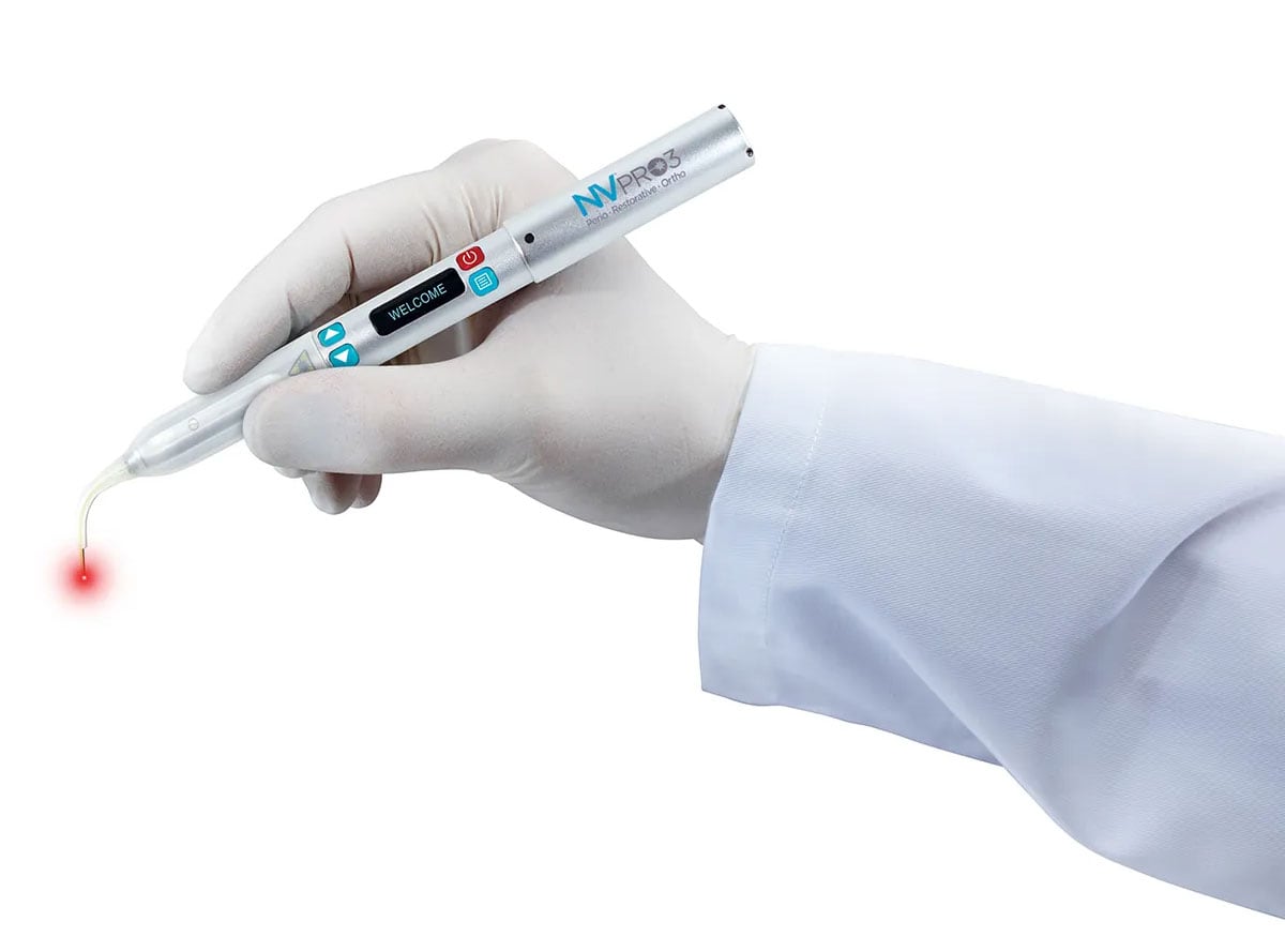
The NV® PRO3 Microlaser has set new quality levels for convenience, portability and ease-of-use among soft-tissue diode dental lasers. Optimized for all of your periodontal, restorative, orthodontic and surgical procedures, the latest evolution in cordless dental diode lasers enable you to deliver the benefits of laser dentistry to each patient, while increasing practice production across all departments.
Digital Radiography
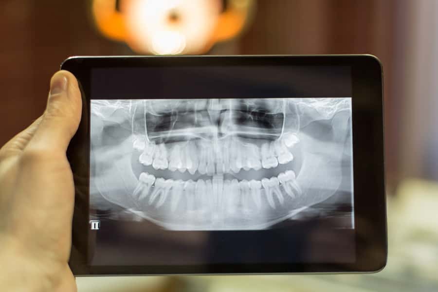
Digital x-rays are the newest technology used in dentistry to take and archive dental x-rays. Digital x-rays significantly reduce the amount of radiation as compared to traditional dental x-rays. This technique captures a digital picture of teeth with their supporting bone structures and stores the images on a computer in our dental office. You and your dentist will be able to instantly view your x-rays and enlarge the image to aid in the identification of dental problems and to gauge your dental health. Your dentist will use this information to create an individualized treatment plan.
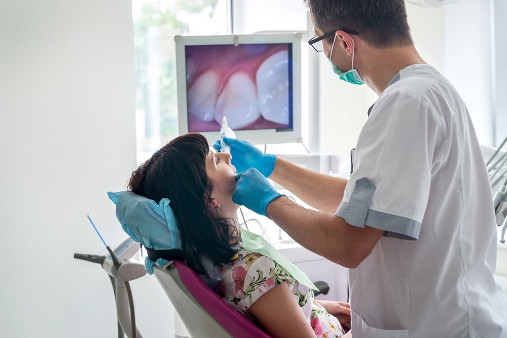
Intra-Oral Camera
Taking pictures of the teeth and supporting tissue with a small video camera about the size of a pen is a wonderful addition to dentistry today. The intra-oral camera creates digital images that can be stored on a computer. The images are shared with our patients so they can join us in the "co-diagnosis" of problems with their dental health. Your dentist can show you how others view your smile and which dental fillings are broken or discolored. We look forward to showing you a dental tour of your mouth in the privacy of your treatment room.